Confocal image of a dorsal root ganglion (DRG) stained with CD31 blood vessels (blue) and TrkA positive expression (magenta).
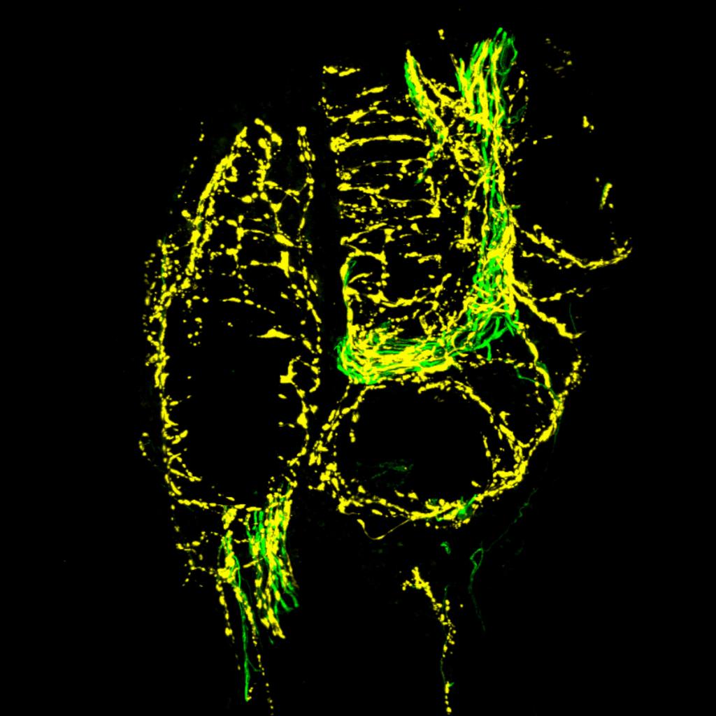
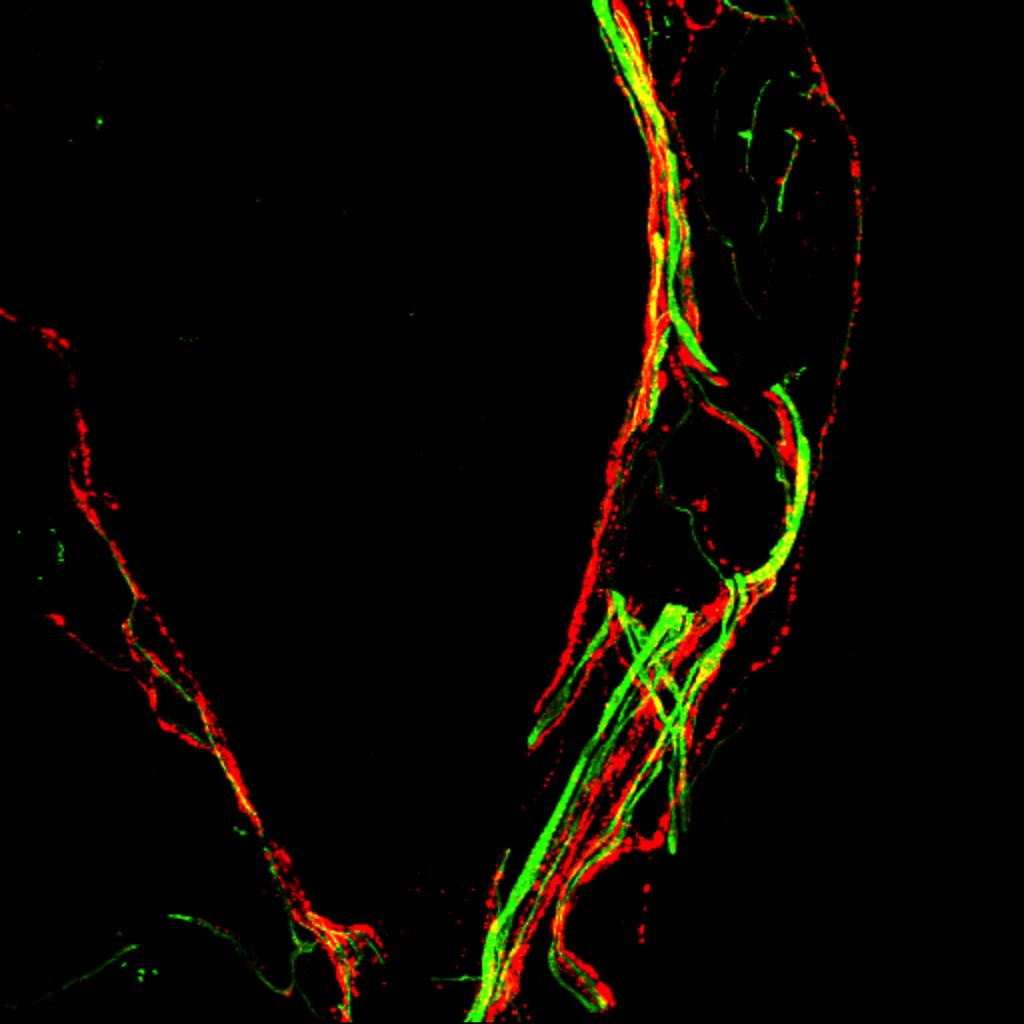
Confocal image illustrating the interaction of NF200 sensory fibers (green) and TH sympathetic nerve fibers (yellow) in a nonhealed, fractured femur. This interaction is never observed in a normal femur.
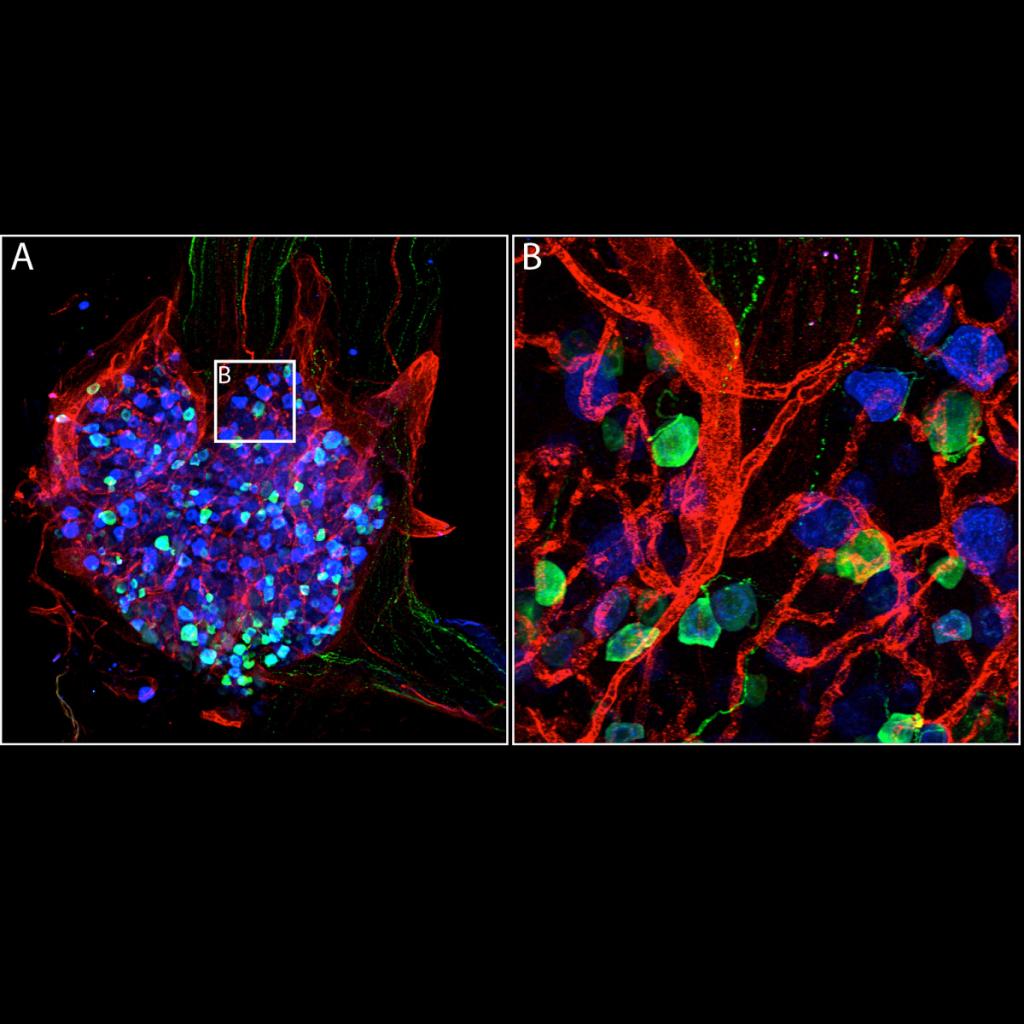
Confocal image of CD31 blood vessels (red), CGRP positive and TrkA positive expression (blue, green) in a dorsal root ganglion. Note the higher power image (B) illustrating the cellular morphology.
Confocal image of a dorsal root ganglion stained for CD31 blood vessels (green) and TrkA positive expression (red).
Confocal image of a neuroma-like structure observed in a nonhealed, fractured femur. Sprouted CGRP (red) and NF200 (green) sensory nerve fibers are observed in a trabecular bone 'pocket' in the bone marrow (blue) adjacent to the initial fracture site.
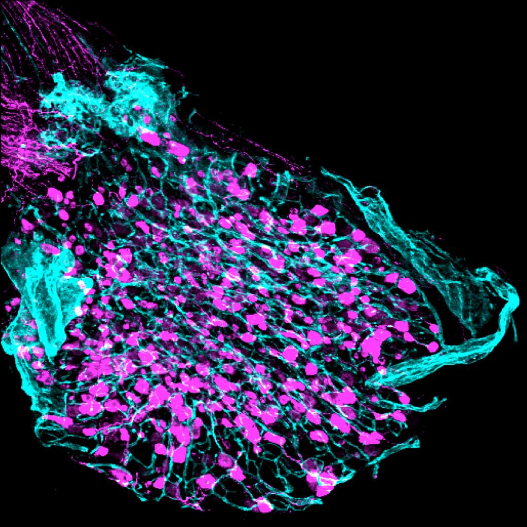
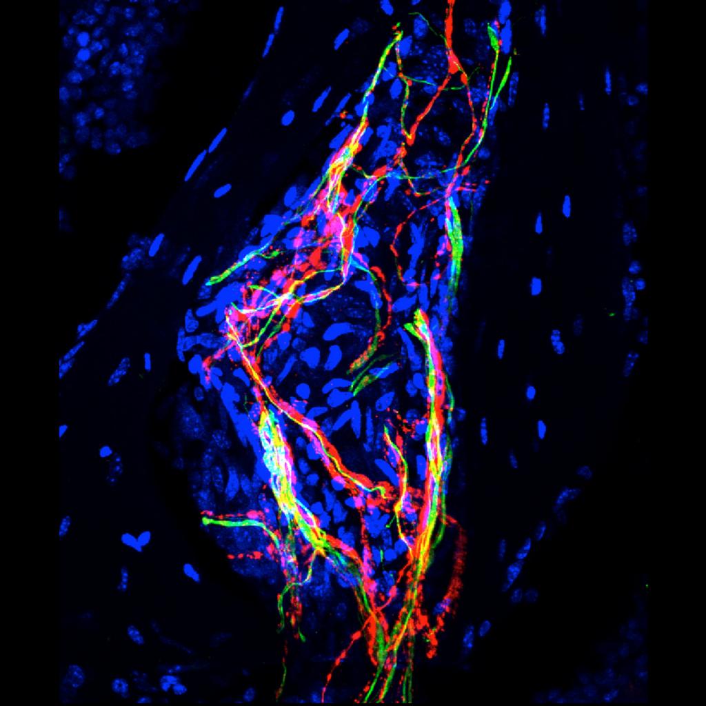
Confocal image of a neuroma-like structure observed in a nonhealed, fractured femur. The structure is comprised of sprouted CGRP (red) and NF200 (green) sensory nerve fibers. These neuroma-like structures are never observed in a normal femur.
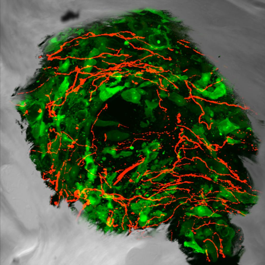
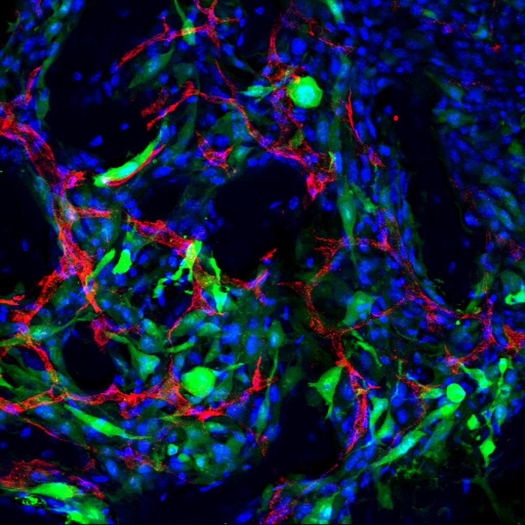
Combined confocal and DIC image of a sensory nerve fibers (red) sprouting into a prostate tumor colony (green). Bone resorbing osteoclasts (yellow) can be seen next to newly formed bone (grey) at the edge of the colony.
Confocal and DIC image of CGRP sensory nerve fibers (red) sprouting into GFP prostate tumor cells (green). Note how the tumor colony is circumscribed by newly woven bone (grey), a hallmark feature of prostate metastasis in bone.
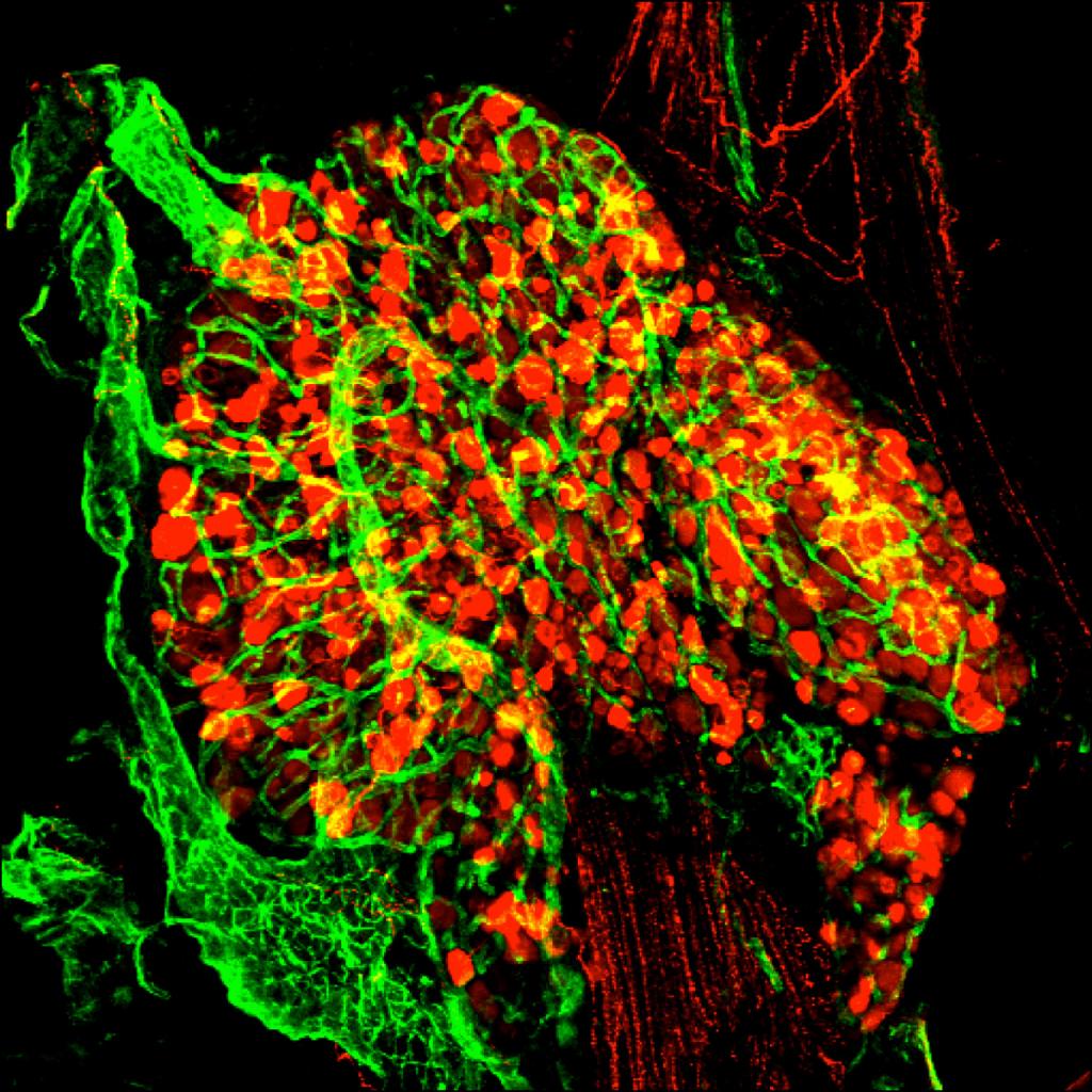
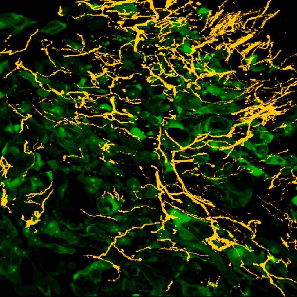
Confocal image of CGRP sensory nerve fibers (yellow) which have sprouted into GFP sarcoma-laden (green) bone marrow. This highly heterogenous nerve growth is never observed in a normal bone.
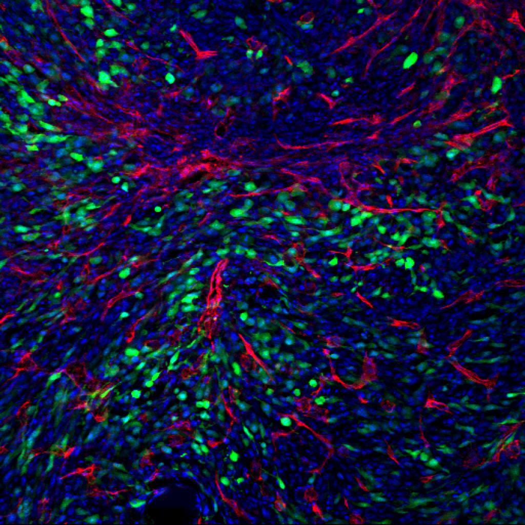
Confocal image illustrating GFP sarcoma tumor cells (green) and CD31 vasculature (red) in the bone marrow (blue).
Confocal image of the bone marrow in a sarcoma-laden femur. Image is stained for DAPI cell nuclei (blue), GFP sarcoma cells (green), and CD31 blood vessels (red).
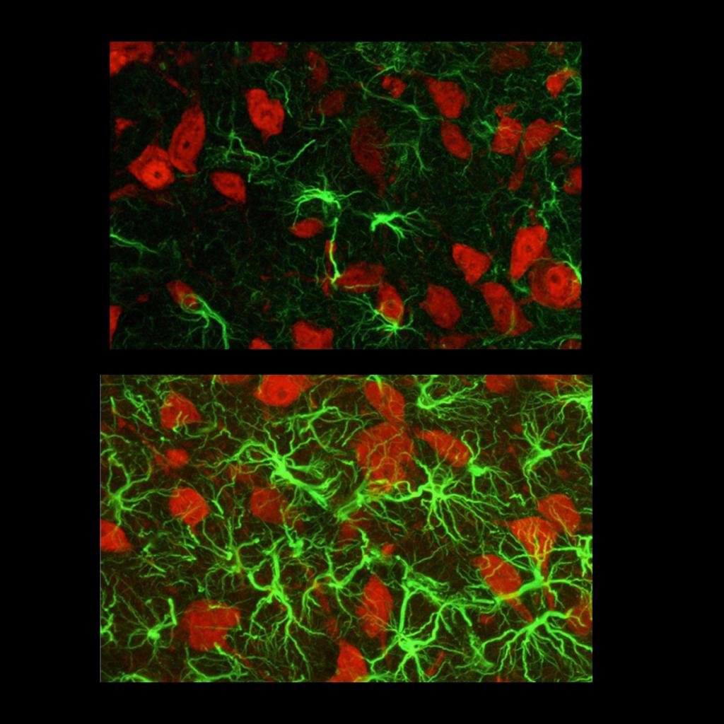
Confocal images from the spinal cord of a normal (top) and sarcoma-injected (bottom) animal. Images are stained with ATF-3, a transcription factor marking cellular damage (red) and NeuN, a marker of neuronal nuclei (green).
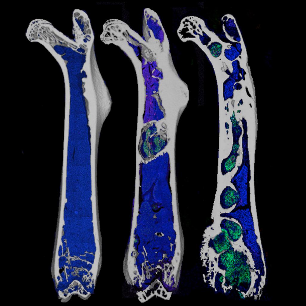
Micro CT images of a femur at early (left), middle (center), and late (right) stages of prostate tumor growth. Each micro CT image was overlaid with a fluorescent image illustrating normal DAPI cell nuclei (blue) and GFP prostate tumor cells (green). Note the increased quantity of prostate tumor colonies with time.
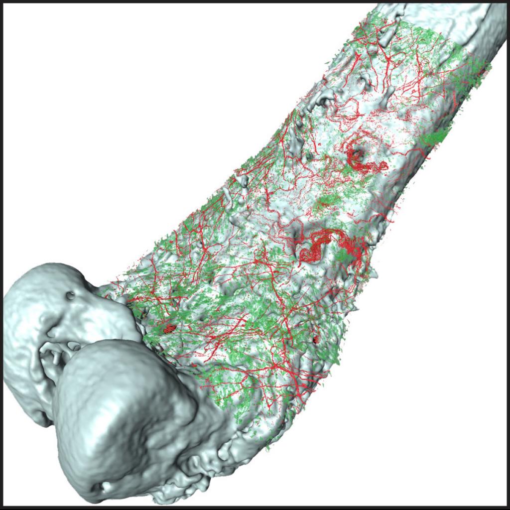
Micro CT image of a femur with a 3D rendering overlay of sprouted CGRP sensory nerve fibers (red) and sarcoma tumor cells (green).